CT technologists at the Drumheller Health Centre had a one in a million experience in December as they scanned their oldest patient to date, a 75 million years old fossilized piece of bone.
The specimen was brought over by The Royal Tyrrell Museum to be scanned using CT technology which gives cross sectional images and the data they acquired was instrumental in revealing a very unusual find.
Over a year ago, work started at the Tyrrell Museum on a new fossilized horned dinosaur.
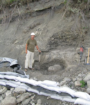
The fossil was found in 2007 when a geologist who was working in the South West part of the province along Old Man river, saw a snout poking out from some rocks.
Darren Tanke, technician at the Royal Tyrrell Museum, then started excavation and collected part of the specimen.
Excavation was stopped when there were concerns about the stability of the hill side, resuming in 2008.
So far a whole skull, with no lower jaw, and a neck frill were recovered, the whole piece being about two meters long.
Don Henderson, curator of dinosaurs at the Royal Tyrrell Museum explains they collected the important sections and how significant frills are: “from the neck back all those horned dinosaurs are very similar. The only way you can tell them apart and the only way they would have told themselves apart and recognized their own kind is via frills and the horn on the nose, bump above the eyes.”
Henderson explains that generally a frill is partly a bone that grows out of the skull but there are also bones in the skin that fuse on to that skull as the animal gets older.
When Darren Tanke started to work on the fossil, he noticed some strange patterns on the frill and wanted to ascertain whether these bumps were part of the original skull or whether they were bones from the skin that had fused on.
Such a specimen being quite rare, they wanted to avoid cutting it open so as not to lose even a few millimeters of bone.
“If the rock is the right kind and the cement holding it together is OK, we can peer inside using a CT machine, it doesn’t always work, a lot depends on the type of fossilization,” explained Henderson.
“We would normally take it to Calgary as they have a bigger machine and a permanent technician but we were able to use the CT machine in Drumheller”.
During a period when no patients had been booked, and on the understanding that should an emergency occur the project would need to be postponed, CT technologists at The Drumheller Health Centre figured out the best techniques on how to scan this unusual item.
Henderson and Tanke took two CDs with them, one containing photos with an image viewer and another one with DICOM images.
On reviewing the CT data, Henderson and Tanke were surprised by what they saw, “we wanted to see how these bones were attached. If we could see a suture then we would know they were originally separate and they had grown together. We were expecting just a simple contact between them but it turns out one of the bones looked like it was clasping the small bone that it was originally separated from, it is growing around both top and bottom of the skull bones, normally you’d expect just a simple flat contact and it is easy to track its growth, but now we see from using the CT data that it is actually a much more complicated way of growing, the contact between them is much more developed, it really is tightly fastened on.”
This was the first scan taken on this find and the research is still at the very early stages, expected to be completed within the next six months, “we haven’t received the final answer on this...we are waiting to see if there’s going to be any more opportunities for putting pieces in the CT machine” explained Henderson.
The name of this specimen? “It is so new, it hasn’t got a name yet,” confirmed Henderson.